Normal_barium_swallow_animation_fluoroscopic image
About[]
Fluoroscopy is an imaging technique that uses X-rays to obtain real-time moving images of the internal structures of a patient through the use of a fluoroscope. In its simplest form, a fluoroscope consists of an X-ray source and fluorescent screen between which a patient is placed. However, modern fluoroscopes couple the screen to an X-ray image intensifier and Charge-coupled device (CCD) video camera allowing the images to be recorded and played on a monitor.
The use of X-rays, a form of ionizing radiation, requires the potential risks from a procedure to be carefully balanced with the benefits of the procedure to the patient. While physicians always try to use low dose rates during fluoroscopic procedures, the length of a typical procedure often results in a relatively high absorbed dose to the patient. Recent advances include the digitization of the images captured and flat panel detector systems which reduce the radiation dose to the patient still further.[1]

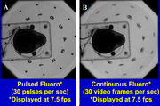
A. Pulsed versus B. continuous fluoro mode
Usage[]
Fluoroscopy produces real-time motion such as continuous, pulsed, or high dose (greater than 20R/min) scanned images of internal structures of the body in a similar fashion to radiography, but employs a constant input of x-rays, at a lower dose rate. Contrast media, such as barium, iodine, and air are used to visualize internal organs as they work. Fluoroscopy is also used in image-guided procedures when constant feedback during a procedure is required. An image receptor is required to convert the radiation into an image after it has passed through the area of interest. Early on this was a fluorescing screen, which gave way to an Image Amplifier (IA) which was a large vacuum tube that had the receiving end coated with cesium iodide, and a mirror at the opposite end. Eventually the mirror was replaced with a TV camera.
Mostly, fluoroscopic tubes are considered under the table tubes because the tube head is literally under-the table.
Regulatory standards[]
Refer to 21 CFR, SUBCHAPTER J, Sec. 1020.32 Fluoroscopic equipment.[2]
Reference[]
- ↑ FDA. "Medical Imaging." 06/05/2014. http://www.fda.gov/Radiation-EmittingProducts/RadiationEmittingProductsandProcedures/MedicalImaging/ucm2005914.htm
- ↑ 21 CFR, SUBCHAPTER J--RADIOLOGICAL HEALTH, PART 1020 -- PERFORMANCE STANDARDS FOR IONIZING RADIATION EMITTING PRODUCTS, Sec. 1020.32 Fluoroscopic equipment.http://www.accessdata.fda.gov/scripts/cdrh/cfdocs/cfcfr/CFRSearch.cfm?FR=1020.32